
Tiny programs run inside our mouths, shaping roots, bone, and bite. New work maps those programs back to stem cells, the living engines behind future regenerative dentistry. Scientists from Japan and the United States traced cell lineages and signals that decide whether young tissues become root, ligament, or jaw bone, while they outline a practical path to restore lost structures that drills, dentures, and implants still fail to mimic. The mystery begins at the growing tooth’s tip.
Why true tooth regeneration remains so difficult
Teeth are not individual components. They are ecosystems. Crowns, roots, pulp, ligament, and alveolar bone develop together during growth, communicating continuities of internal messaging that coordinate time and form. As each piece of tooth tissue depends upon the maturation of other components at varied rates, the patient treatment plan must also sequence time and choreography. It is not just about replacing a visible piece or one surface that is now missing.
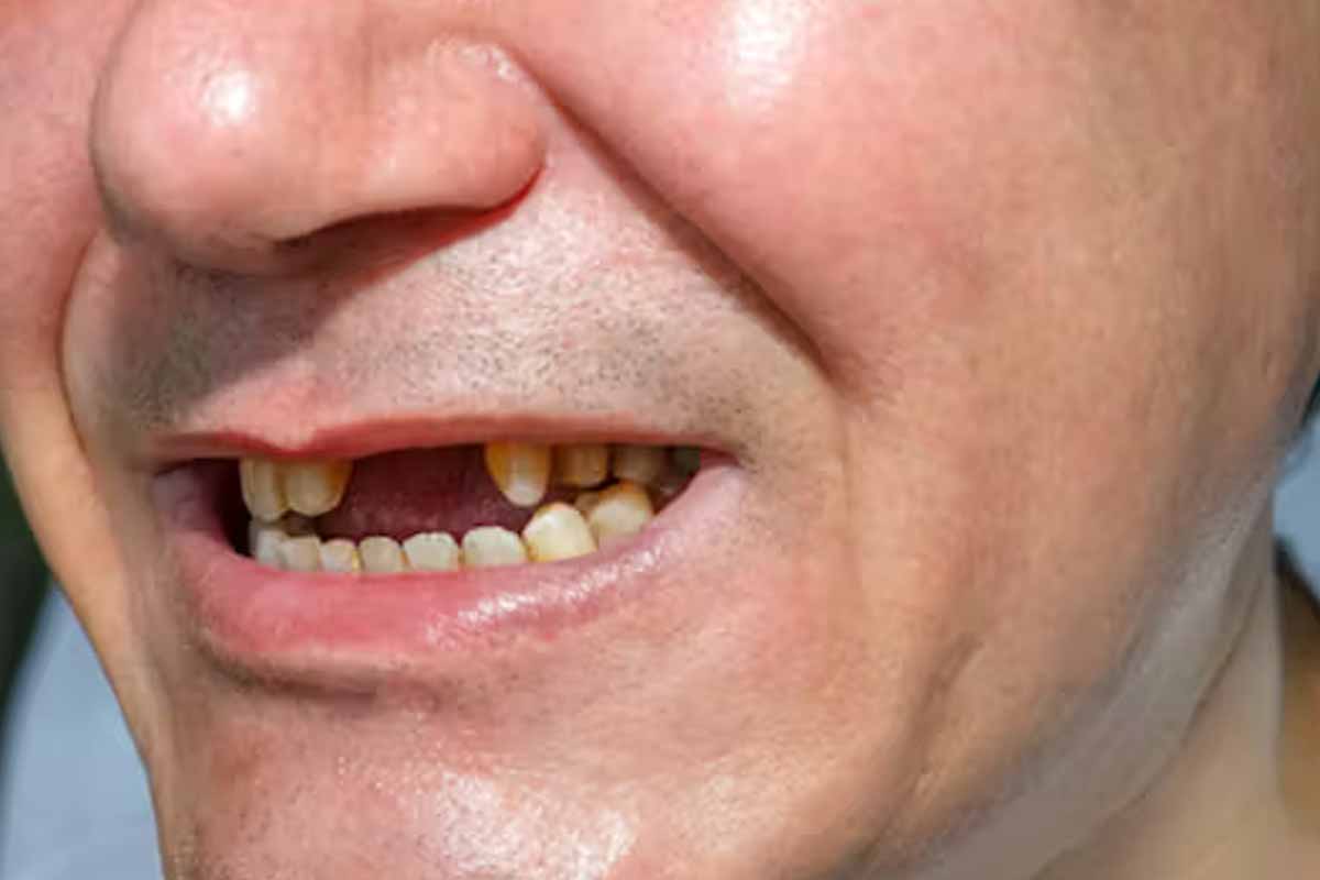
Dentures and titanium implants restore chewing and smile, yet they still miss the natural feel and biological feedback of a living root. The jaw senses load through the periodontal ligament; without that loop, bone remodels differently over years, and long-term integration can drift in subtle ways from nature.
Researchers therefore study the body’s own builders, stem cells, to recreate that integration. Signals from enamel organ, pulp, and jawbone must align so young cells choose odontoblast, cementoblast, or osteoblast fates with precision. Mapping those choices, and when they lock in, depends on timing maps and controllable molecular switches.
How stem cells decisions are tracked and steered
An international team led by Assistant Professor Mizuki Nagata at Science Tokyo, with collaborators at UTHealth and the University of Michigan, traced cell fates during root growth. They focused on the apical region, the growing tip where choices harden. There, root, ligament, and supporting bone diverge from shared beginnings.
Using genetically modified mice and lineage tracing, the team labeled emerging populations with fluorescent tags, then silenced specific genes to test causality. Microscopy linked signal pulses to fate, while pathway inhibitors clarified which cues push cells toward root, cementum, or alveolar bone under carefully controlled regenerative conditions.
The framework ties canonical Wnt activity to root elongation decisions, and relates Hedgehog–Foxf suppression to bone commitment. Because stem cells interpret these signals in real time, interventions must be timed and dosed, not merely switched on. That insight turns vague hope into a plan for controllable regeneration.
From lab maps to clinical possibilities
The work organizes tooth development into programmable steps. With a clearer map, dentists and surgeons could one day seed specific cell types into damaged sites, while biomaterials deliver timed signals. That approach aims for living roots that talk to bone, not just artificial anchors that sit in it.
Because periodontal ligament feedback preserves bone, designs that restore ligament fibers may reduce long-term resorption around replacements. Targeted therapies might also rescue threatened teeth by re-building root tissues before extraction becomes inevitable, so fewer patients cycle between temporary fixes and major reconstructions. Stepwise goals keep progress credible.
Therapies will rely on curated stem cells and synchronized cues. Safety testing, dose windows, and immune responses remain key checkpoints. A mechanistic framework lets trials measure the right endpoints, root length, cementum quality, and ligament function over time, because success depends on whole-organ performance in patients, not one metric.
Two stem cells lineages shape roots and jaw support
The team identified mesenchymal populations with distinct futures. One arises in the apical papilla within the epithelial root sheath. These CXCL12-expressing cells sit at the root’s growing tip and, under canonical Wnt signals, choose odontoblast fate for dentin or switch to cementoblast programs along the elongating root surface.
Under regenerative conditions, the same apical lineage can generate osteoblasts that contribute to alveolar bone. That plasticity links root extension with socket repair in vivo. A second lineage concentrates in the dental follicle, where PTHrP-expressing cells diversify into cementoblasts, periodontal ligament fibroblasts, and bone-forming osteoblasts under context-dependent cues.
Crucially, their alveolar bone destiny depends on turning down Hedgehog–Foxf activity. Nagata’s group shows that suppressed Hedgehog signaling unlocks that path, revealing a tooth-specific mechanism for bone formation. Because stem cells read these programs locally, delivery systems must aim signals to the apical and follicle niches with precision.
Dates, methods, and signals that define a research roadmap
Two companion papers in Nature Communications, Volume 16, appeared July 1 and July 2, 2025. They report coordinated studies from Science Tokyo, UTHealth, and the University of Michigan. Together they align lineage-tracing readouts with pathway perturbations, so findings flow from cause-and-effect tests and the logic remains reproducible.
Signals carry the action. Canonical Wnt correlates with root elongation programs. Hedgehog–Foxf suppression unlocks alveolar bone commitment in the follicle lineage. The apical papilla and follicle occupy tight niches. Dose, timing, and delivery vectors will decide whether tissues rebuild harmoniously or drift from intended patterns.
With better control knobs for stem cells, developers can design staged interventions: first stabilize the site, then re-establish ligament, and finally thicken bone. Preclinical models in mice provide proof of mechanism, and future work will translate those steps into human protocols with clear milestones and decision gates for clinicians.
What today’s map suggests about tomorrow’s tooth repair
Regeneration moves when models turn into instructions and clinics can follow them. These studies show where choices are made, who makes them, and how to nudge them safely. By linking signals to outcomes at the root tip and follicle, researchers give dentistry a playbook. With carefully timed cues and living stem cells, replacements may one day feel native, integrate, and endure—teeth that grow, anchor, and adapt again. Cautious steps now build the foundation for durable change.